 Follow Us on Google News
 Follow Us on Google Discover
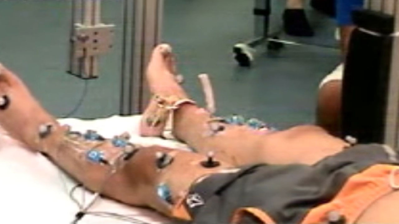
*Sterilised silk from moths could repair damage by producing material which acts as a ‘scaffold’
A research participant at the University of Louisville, United States (U.S.), with a complete spinal cord injury, who had lost motor function below the level of the injury, has regained the ability to move his legs voluntarily and stand six years after his injury.
A study published today in Scientific Reports describes the recovery of motor function in a research participant who previously had received long-term activity-based training along with spinal cord epidural stimulation (scES). In the article, senior author Susan Harkema, Ph.D., professor and associate director of the Kentucky Spinal Cord Injury Research Center (KSCIRC) at the University of Louisville, and her colleagues report that over the course of 34.5 months following the original training, the participant recovered substantial voluntary lower-limb motor control and the ability to stand independently without the use of scES.
Previous research at KSCIRC involving four participants with chronic clinically motor-complete spinal cord injury found that activity-based training with the use of scES — electrical signals delivered to motor neurons in the spine by an implanted device — allowed the participants to stand and to perform relatively fine voluntary lower limb movements when the scES device was activated. Andrew Meas was one of the four participants in that study.
[ad]
The original training protocol included daily one-hour activity-based training sessions with the aid of epidural stimulation. During these sessions, the participant trained on standing activity for several months, followed by several months of training on stepping. After completing a nine-month training program in the lab, Meas continued activity-based stand training at home. After a year of independent training, he returned to the lab to train for three months in a revised activity-based training schedule. The revised training called for two daily one-hour training sessions and included both stand and step training each day, all with the aid of epidural stimulation.
Also, scientists claim silk could help repair damaged spinal cords by producing material which acts as a ‘scaffold’. A team of British researchers found cleaned, sterilised silk from a breed of moths had properties well suited to spinal repair.
A modified version of the lightweight material could support nerve growth across damaged regions, they believe.
Dr. Fritz Vollrath, from Oxford University, said the findings are the ‘most important and exciting’ conducted on the values of silk yet.
Both he and a team of researchers at Aberdeen University, believe derivatives of silk can aid natural regeneration for major spinal injuries.
There is currently no cure for serious traumas because the nerves can’t cross the scar tissue barrier and the cavity forming in the column after the injury.
The team discovered that modified Antheraea pernyi silk had important properties desirable in a ‘scaffold’ suitable for spinal repair.
Their findings, published in the journal Scientific Reports, were derived from petri dish tests on rat cells.
It has the correct rigidity, as if it is too rigid it can harm the surrounding spinal cord tissue but if it is too soft the nerves would fail to grow across it.
The AP silk has a chemical sequence on its surface that binds to receptors on the nerve cells, encouraging them to attach to the material and grow along it.
Additionally, it didn’t trigger a response by the immune system cells that would be present in the spinal cord, therefore minimising inflammation.
Finally, the AP silk degrades gradually over time. This means, after it has supported the early growth of nerves across the injury site the material dissolves.
These nerves then take over the role as scaffold, supporting further nerve growth in the sight of injury.