 Follow Us on Google News
 Follow Us on Google Discover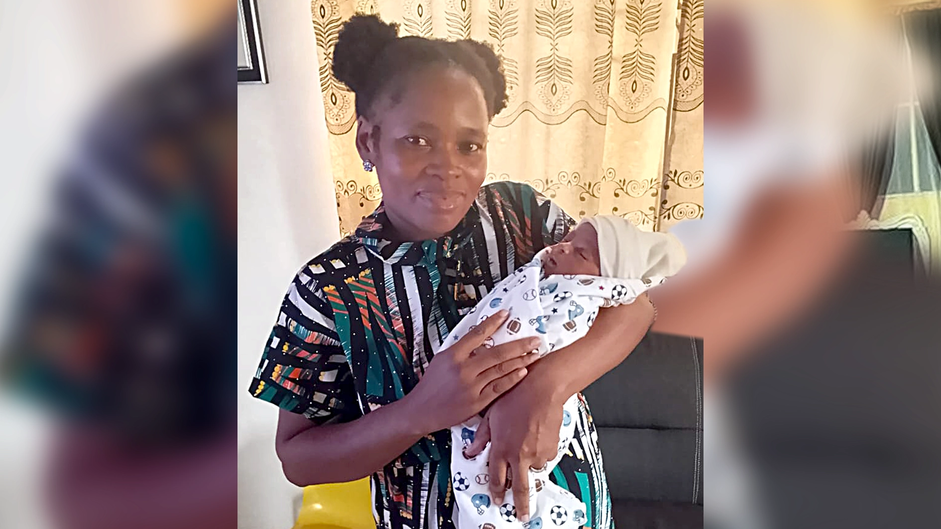
• ‘Scan showed nothing… until doctors found my Baby outside the womb’
• Inside LUTH’s extraordinary efforts to save mother, baby
• Why abdominal pregnancies remain one of obstetrics’ rarest emergencies
• How months of monitoring, teamwork and preparation changed the outcome
For Michelle Olarinde, the journey to motherhood had already been long before she conceived. After years of waiting and hoping for a child, she became pregnant and expected a routine pregnancy like every other expectant mother.
Instead, she found herself at the centre of one of the rarest forms of pregnancies: one that would keep her in hospital for three months and require a multidisciplinary team of specialists to monitor her until doctors considered it safe to deliver her baby.
Recalling how it all began, Olarinde said she discovered she was pregnant after missing her menstrual period on November 8, 2025. A home pregnancy test confirmed her suspicion and she immediately informed her physician, Dr. Babatunde Bamidele, who advised her to wait another two weeks before going for an ultrasound scan.
The result of the first scan was unexpected. It showed no pregnancy inside her womb. Believing it might have been too early for the pregnancy to appear, doctors advised her to return after another two weeks. She did, but the second scan produced the same result. A blood test was then requested and it confirmed she was pregnant. However, another scan still failed to locate the pregnancy.
Olarinde said the conflicting results became increasingly difficult to understand because her body was already showing signs of pregnancy.

“When I went for the scan, it was showing there was nothing in the womb, so, I was advised to wait another two weeks because they felt the baby might still be too young to show. I waited, repeated the scan and it was still showing nothing. I informed Dr. Bamidele again and he asked me to do a blood test. The blood test showed I was pregnant, but another scan still showed nothing.
“I was worried because the blood test was positive and I was already having pregnancy symptoms like vomiting, headaches and all that, yet the scans kept showing nothing. I knew there was a pregnancy because I could feel the symptoms,” she said.
Unable to explain the conflicting findings, Michelle said she prayed for God to reveal what doctors had not been able to see.
“I prayed one prayer. I said, ‘God, the doctors are not seeing anything in my body. The next scan I do, show them something.’ I was already feeling movement and my stomach was paining me somehow. I knew God had done something, but I did not know the baby was outside the womb.”
Her prayer was answered during another examination.
A more detailed scan revealed that the pregnancy was not inside the uterus but growing in her abdomen, a rare and potentially life-threatening condition known as an abdominal pregnancy. By then, the pregnancy had already reached 20 weeks.
Olarinde said her doctor immediately referred her to the Lagos University Teaching Hospital (LUTH) for specialist care. When the doctor carried out another thorough scan, he found out that the baby was actually there but not in the womb. It was outside the womb and the pregnancy was already 20 weeks.
She was admitted to LUTH on April 8, 2026. Rather than discharge her, doctors explained that because of the rarity of the pregnancy, she would remain in hospital under close observation.
Their plan was to monitor both mother and baby closely and intervene only if continuing the pregnancy became too dangerous.
Week after week, the pregnancy progressed without the complications doctors feared. At every milestone, the medical team reassessed the situation, balancing the risks to her against the baby’s chances of survival.
“They said if there was danger at 24 weeks, they would remove the pregnancy to save my life. When I got to 24 weeks, there was no danger, so they continued monitoring me. At 27 weeks, they said if there was any danger, they would remove the baby and put the baby in the incubator. Nothing happened. At 30 weeks, I was still strong. At 32 weeks, it was the same. Then, at 34 weeks, anytime the baby moved, I started having serious pains and I was rushed to the theatre.
“All through my stay in LUTH, Dr. Opeyemi Akinajo and all the other doctors really took good care of me. I give them a plus.”
Despite being told she was carrying a high-risk pregnancy, she said fear never overwhelmed her because she believed God had a purpose for the pregnancy.
By the time doctors discovered the baby was growing outside her womb, she said the pregnancy had already advanced to 20 weeks, deepening her resolve to continue.
She also credited her husband for standing by her throughout the difficult months. “My husband encouraged me throughout.
I just kept trusting God because I believed God started it and He would complete it.”
As the date for surgery approached, Olarinde said she remained confident that both she and her baby would survive despite repeatedly hearing doctors describe the pregnancy as high-risk.
Seeing the number of consultants, doctors and nurses gathered before the operation also strengthened her confidence.
“Before the surgery, I just had this confidence that my baby and I would come out alive because I believed God would complete what He had started.
“I remember seeing many doctors, consultants and nurses fully on ground. Even doctors I did not know came around before the surgery. That gave me joy and confidence.”
The operation was successful although she was still recovering from the anaesthesia, hearing her baby’s first cry, she knew the months of uncertainty had finally come to an end.
“When I heard my baby cry after the surgery, I just said, ‘Thank you, Jesus. We made it’.”
Looking back on the experience, she told The Guardian that her faith sustained her through the uncertainty, while acknowledging the emotional and financial burden of spending almost three months in hospital.
“I want people to understand that trusting God was what kept me going. There were difficult moments, but I kept holding on to God because I believed He would see me through.
“I stayed in the hospital for almost three months, from April to July. There were hospital bills, drugs and many other expenses, but I just saw them as part of the journey because I was grateful that both my baby and I came out alive.”
One Of Obstetrics’ Rarest Emergencies And Risks That Come With It
ITS rarity is reflected in the numbers. Advanced abdominal pregnancy occurs in about one out of every 10,000 to 30,000 pregnancies and accounts for about one per cent of ectopic pregnancies worldwide.
An ectopic pregnancy is a dangerous medical condition where a fertilised egg implants outside the uterus, most often in a fallopian tube.
It cannot survive and can cause severe internal bleeding if the tube bursts.
During this period, sharp or crampy pain is felt in the lower belly or pelvis. Also, there is always light or heavy unusual vaginal bleeding, making the woman dizzy, faint, or feeling very weak.
Although uncommon, it remains one of the most dangerous forms of pregnancy because the placenta may attach itself to organs, ligaments and blood vessels that are not designed to support pregnancy.
For women who survive long enough for the pregnancy to advance, the greatest danger is often not the delivery of the baby but the management of the placenta.
Removing it can result in severe bleeding, while leaving it behind may expose the mother to complications after delivery.
Fetal mortality in abdominal pregnancies has been reported between 40 per cent and 90 per cent, making successful outcomes involving both mother and child extremely uncommon. That was the challenge awaiting the specialists at LUTH.
Months Of Monitoring, Planning, Preparing For The Unexpected
THE lead consultant obstetrician and gynaecologist, Dr Akinajo, said the greatest challenge was not delivering the baby but managing the placenta.
She said though abdominal pregnancies are rare, the placental implantation site largely determines the clinical outcome, as attachment to major blood vessels or vital abdominal organs can significantly complicate management.
Attempting to remove it in such circumstances may trigger catastrophic haemorrhage.
Describing the case as one of the most challenging pregnancies she has managed, Akinajo said the patient was referred to LUTH at about 21 weeks’ gestation after repeated scans at the fertility centre suggested the pregnancy was developing outside the uterus.
On arrival, she said, another ultrasound examination carried out at LUTH confirmed the diagnosis of an advanced extra-uterine pregnancy.
The medical team immediately counselled the patient and her husband on the risks associated with continuing the pregnancy, including severe haemorrhage, fetal loss, injury to surrounding organs and the possibility that the baby could develop deformities resulting from mechanical compression.
Despite the risks, the patient expressed a strong desire to continue the pregnancy after years of infertility, prompting doctors to admit her for continuous monitoring rather than attempt immediate intervention.
According to her, the hospital immediately activated a multidisciplinary management plan involving obstetricians, anaesthetists, neonatologists, haematologists, radiologists, general surgeons, cardiothoracic surgeons and urologists.
She explained that each specialist played a critical role in her management because the complexity of the pregnancy depended largely on the placental implantation site and the surrounding structures involved, which required careful preoperative assessment and multidisciplinary surgical planning.
An MRI later revealed that the placenta was attached to the upper part of the uterus, extended towards the left fallopian tube and had dense adhesions involving the sigmoid colon and surrounding tissues. The imaging also showed that it was lying close to major abdominal blood vessels, raising concerns that delivery could result in life-threatening bleeding.
“The placenta was what worried us most, we knew that if it had invaded major blood vessels or other vital structures, removing it could become extremely dangerous.”
To prepare for every possible outcome, she said blood donation was arranged in advance, emergency blood transfusion protocols were activated and the patient’s anaemia was corrected with intravenous iron therapy to improve her haemoglobin level before surgery.
The baby also received antenatal support through medications administered to the mother to accelerate lung maturity in anticipation of possible premature delivery.
Throughout the admission, specialists met repeatedly to review the patient’s condition, reassess the risks and refine their surgical strategy.
According to her, the team prepared for every possible scenario, including the need for intensive care, emergency bowel surgery, urological reconstruction and massive blood transfusion if severe bleeding occurred.
As the pregnancy approached 34 weeks, the patient developed worsening abdominal pain, particularly whenever the baby moved.
She also began vomiting and struggled to tolerate meals, prompting doctors to conclude that waiting any longer could increase the danger to both mother and child.
The multidisciplinary team reconvened and decided to move the operation forward.
Operation To Save Both Mother, Baby
ON the day of surgery, the multidisciplinary team of consultant specialists assembled in the operating theatre, while blood products, specialised equipment, and additional emergency support teams were kept on standby.
After opening the abdomen, the surgeons identified a live male baby lying within its gestational sac in the abdominal cavity. The baby was delivered successfully, cried almost immediately after birth, and handed over to the neonatal team for assessment.
Attention then shifted to what doctors had feared most the placenta.
Fortunately, surgeons found that though it was firmly attached to the uterus, sigmoid colon and surrounding tissues, careful dissection allowed it to be removed completely without causing the catastrophic bleeding they had anticipated.
Although the patient required blood transfusion during the procedure, the blood loss remained within manageable limits, and there was no injury to the bowel, urinary tract or other adjacent abdominal organs.
Equally reassuring, the baby showed no evidence of structural abnormalities, despite the increased risk of fetal deformation and other complications associated with an abdominal pregnancy.
The mother recovered without requiring admission into the intensive care unit, while both mother and baby progressed well enough to be discharged home.
Reflecting on the outcome, Akinajo attributed the success to months of meticulous planning, constant monitoring and collaboration with other specialties.
She said no single doctor could have managed the pregnancy alone, noting that the willingness of every specialist to work together, combined with the patient’s remarkable cooperation throughout her admission, proved critical to the successful outcome.
She added that every completed pregnancy should be managed according to its unique circumstances rather than a fixed protocol, stressing that multidisciplinary care remains essential whenever rare, high-risk conditions are encountered.
For the medical team, the successful delivery represents more than a rare clinical achievement.
It is expected to be documented in medical literature, contributing to the limited global evidence on advanced abdominal pregnancies in which both mother and baby survive.
‘We Trusted The Doctors, Held On To Hope’
OLARINDE’S husband told The Guardian that the diagnosis also came as a shock to him, following the couple’s years-long journey through infertility and IVF treatment.
He recalled that after repeated pregnancy tests confirmed conception but ultrasound scans continued to show an empty uterus, the eventual discovery that the baby was developing outside the womb brought mixed emotions.
While relieved that the pregnancy had finally been located after weeks of uncertainty, he also understood the enormous risks that lay ahead.
“I first thanked God that the baby had finally been seen after two positive pregnancy tests and repeated scans that could not locate it, I encouraged my wife that whatever the doctors advised, we would comply. I knew it wasn’t going to be easy, but we were ready to give it a try, trusting God.”
The months that followed proved physically, emotionally and financially demanding.
With his wife admitted to LUTH for more than three months, he struggled with the reality of leaving her in hospital each day while worrying about the mounting cost of treatment.
“It wasn’t easy leaving her alone in the hospital, the financial aspect was another challenge because I knew managing such a rare pregnancy would require a lot.”
Despite the risks repeatedly explained by doctors, he said neither he nor his wife allowed fear to overwhelm them.
Instead, they focused on following medical advice while holding firmly to their faith, even when relatives expressed concerns about the dangers surrounding the pregnancy.
“There were people who tried to make us afraid, but we refused to dwell on negative comments. We believed God would see us through, and we also resolved to follow everything the doctors asked us to do,” the husband said.
The most anxious moment came during surgery.
After the baby was delivered, he was informed that doctors were still working to stabilise his wife.
“I was told the baby was safe, but they were trying to stabilise my wife, I told one of the doctors that God would perform a miracle through their hands. When they later told me that my wife was also stable, I was overwhelmed with joy. I began singing praises to God.”
Looking back, he described the birth of their son after eight years of marriage as a life-changing experience.
He urged other couples facing complex pregnancies to seek prompt care from qualified specialists rather than delay treatment.
“Our consultant referred us to LUTH immediately the condition was discovered, and that decision made all the difference, the medical team monitored my wife throughout the pregnancy and gave us hope. We are grateful for their professionalism and dedication,” he said. The rarity of the condition meant the patient required care that extended far beyond routine obstetric management.
Why Specialist Care Made The Difference
ACCORDING to the Chairman, Medical Advisory Committee (CMAC) of the Lagos University Teaching Hospital (LUTH), Prof. Ayodeji Oluwole, abdominal pregnancy is one of the rarest forms of ectopic pregnancy and is typically managed only in specialist centres because of the high risk it poses to both mother and baby.
He explained that unlike a normal pregnancy, where the embryo implants within the lining of the uterus, an abdominal pregnancy develops when the embryo implants somewhere inside the abdominal cavity after conception.
In IVF pregnancies, he said, although the embryo is transferred into the uterus, it may in rare instances migrate through the fallopian tube into the abdominal cavity, where it attaches to surrounding tissues and continues to grow.
According to medical literature, ectopic pregnancies occur in about one to two per cent of all pregnancies, while abdominal pregnancies account for roughly one per cent of ectopic pregnancies, making them exceptionally rare.
Maternal mortality associated with abdominal pregnancy has been reported to be several times higher than that of tubal ectopic pregnancies, while fetal mortality ranges between 40 and 90 per cent.
Oluwole said the patient’s fertility specialists recognised the unusual nature of the pregnancy and immediately referred her to LUTH because such cases require facilities and expertise rarely available outside tertiary hospitals.
“When she arrived, she was admitted immediately because the pregnancy could rupture at any time, leading to catastrophic bleeding that could threaten her life,” he said.
Rather than rush into surgery, he explained, the medical team adopted conservative management, carefully monitoring both mother and baby while allowing the pregnancy to progress to a stage where the baby would have a better chance of surviving outside the womb.
According to him, the patient remained on admission for months as doctors closely monitored the pregnancy, knowing that any sudden deterioration could require emergency intervention.
The decision to continue the pregnancy, he noted, was constantly weighed against the possibility of life-threatening complications.
By the time the pregnancy reached 34 weeks, the fetus had attained a level of maturity that significantly improved the chances of survival. However, the patient later developed severe abdominal pain, prompting the medical team to proceed with emergency delivery earlier than initially planned.
Oluwole attributed the successful outcome largely to early planning and collaboration among specialists from multiple disciplines.
He said consultant obstetricians, anaesthetists, paediatricians, urologists, general surgeons, haematologists and other specialists jointly reviewed the case long before surgery, identifying possible complications and preparing responses for each scenario, including the availability of sufficient blood products in anticipation of severe haemorrhage.
The professor described the outcome as one of the rare successes recorded in advanced abdominal pregnancy.
“In over three decades of medical practice, I have only encountered three abdominal pregnancies,” he said, describing the condition as an uncommon occurrence that deserves documentation in medical literature because of its rarity.
He noted that many abdominal pregnancies end before viability following rupture or severe complications, making the successful delivery of a healthy baby at 34 weeks particularly remarkable.
Another unusual aspect of the surgery, according to him, was the successful removal of the placenta.
In many abdominal pregnancies, surgeons are often forced to leave the placenta inside the abdomen because attempts to remove it can trigger uncontrollable bleeding, especially when it is attached to major blood vessels or vital organs.
In this case, however, the placenta was attached to the uterus and surrounding tissues in a manner that allowed surgeons to remove it successfully without the devastating blood loss often associated with such operations.
Beyond the clinical success, Oluwole said the case also highlights the critical role of tertiary healthcare facilities in managing highly specialised medical conditions.
He explained that while primary and secondary healthcare centres remain essential for routine care, rare and complex cases require multidisciplinary teams, advanced imaging, specialist surgeons, intensive care support and blood bank services that are usually available only in teaching hospitals.
According to him, increasing Nigeria’s capacity for specialist healthcare remains essential, particularly as the country’s growing population continues to place enormous pressure on existing tertiary institutions.