 Follow Us on Google News
 Follow Us on Google Discover
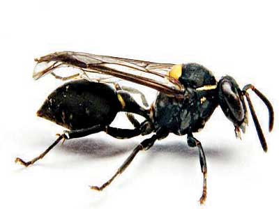 WASPS can be a menace, often found haunting picnics and fruit trees in the summer. However, the venom of one particular breed of wasp is known to contain a potent anticancer ingredient, and now researchers have shown precisely how the venom’s toxin selectively kills cancer cells.
Venom belonging to the Brazilian social wasp Polybia paulista contains the antimicrobial peptide Polybia-MP1 (MP1), which has been demonstrated to inhibit multiple forms of cancerous cells such as prostate cancer, bladder cancer and multidrug-resistant leukemic cells.
Despite this antimicrobial peptide showing great potential as a component of anticancer treatment in humans, researchers have not fully understood exactly how MP1 kills cancer cells.
The new study, published in Biophysical Journal, now reveals how MP1 is capable of killing cancer cells while leaving normal cells unscathed: by attacking lipids on the surface of cancer cells and creating holes that allow important cell molecules to leak out.
“Cancer therapies that attack the lipid composition of the cell membrane would be an entirely new class of anticancer drugs,” explains study co-senior author Paul Beales of the University of Leeds in the United Kingdom (UK).
“This could be useful in developing new combination therapies,” he adds, “where multiple drugs are used simultaneously to treat a cancer by attacking different parts of the cancer cells at the same time.”
The researchers hypothesized that the mechanism behind MP1’s effectiveness against cancer cells would involve the way that cancer cell membranes differ from healthy cell membranes.
One major difference is the positioning of two lipids that form part of the cell membrane: phosphatidylserine (PS) and phosphatidylethanolamine (PE). In cancer cells, PS and PE are located in the outer cell membrane facing outward from the cell, while in healthy cells, they are situated in the inner membrane and face inward.
To test their hypothesis, the researchers created some model cell membranes. Some of these contained PS, some contained PE and some contained both. They then exposed their model membranes to MP1 and observed what happened.
Using a combination of membrane permeability assays and imaging techniques, the researchers revealed that PS increased the binding of the antimicrobial peptide to the cell membrane, while the presence of PE boosted MP1’s ability to quickly disrupt the membrane and increase the size of any holes in it.